

Research and Publications
MRI Features and Pain in First MTP Joint (Big Toe) Osteoarthritis
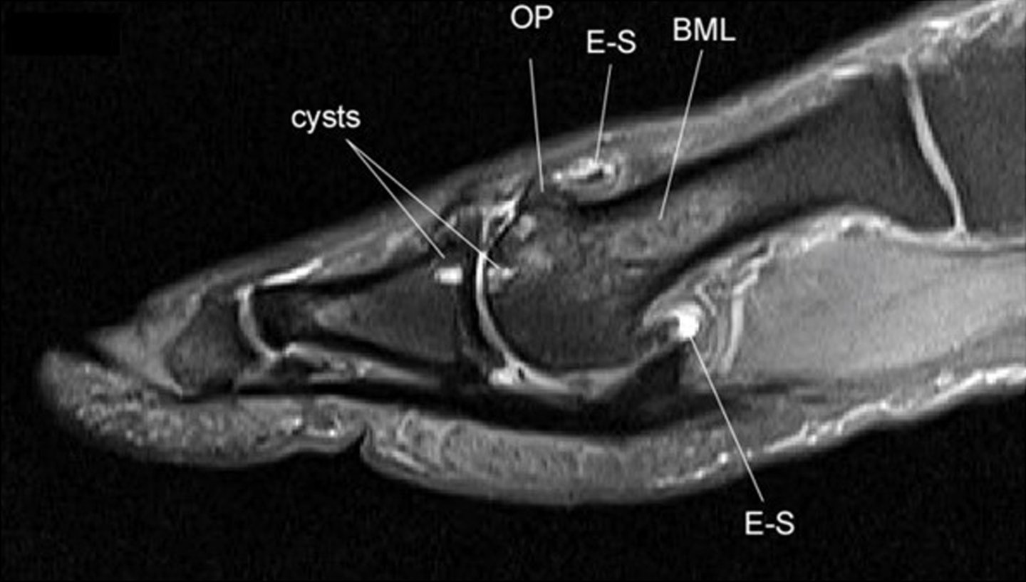
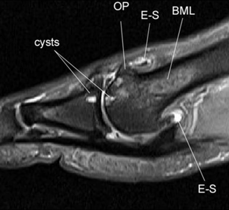
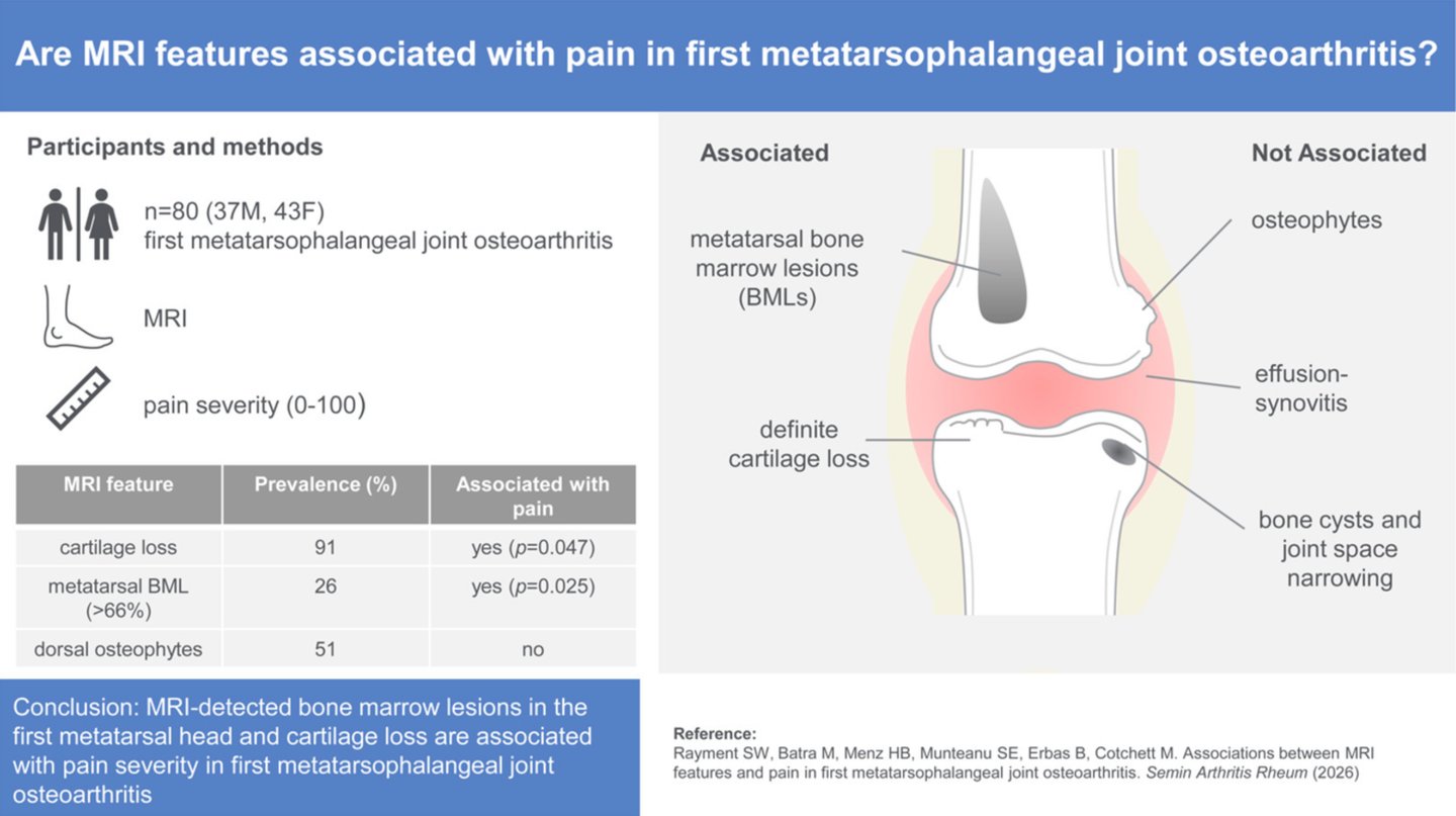
We looked at which MRI-detected structural features in first MTP joint (big toe) osteoarthritis (OA) track with pain severity. Pooling baseline data from 80 participants across two RCTs and utilising standardised atlas features (osteophytes, BMLs, cysts, effusion-synovitis, cartilage loss, JSN).
Osteophytes, JSN, BMLs in the proximal phalanx and sesamoids, cysts, and effusion-synovitis were not associated with pain. What did? First metatarsal head BMLs involving >66% of the head (adjusted p=0.025, stepwise p=0.039) and cartilage loss (stepwise p=0.047).
Which is a bit of a problem for how we currently grade hallux limitus/rigidus severity. Suggesting that JSN and osteophyte size on x-ray correlate poorly with what the patient's actually reporting clinically.
Cross-sectional design, so we can't speak to causality or temporal sequence yet. But it is a reasonable first step and points toward BMLs as a possible imaging biomarker and management strategy for painful OA at the 1st MTPJ.
Results vary based on individual presentation. This is a cross-sectional study, so it identifies associations, not cause and effect, further research is needed to confirm the relationship over time.
Rayment SW, Batra M, Menz HB, Munteanu SE, Erbas B, Cotchett M. Associations between MRI features and pain in first metatarsophalangeal joint osteoarthritis. Semin Arthritis Rheum. 2026 Jun 18;80:153029. doi: 10.1016/j.semarthrit.2026.153029
Scott Rayment is the founder of DynamicStep and a Sports Podiatrist specialising in ankle instability.